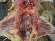
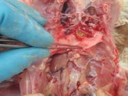
9. Cut the base of the oesphagus
Sever the oesophagus just cranial to liver. Grasp the oesophagus firmly then reflect liver/gut caudally, cutting the mesentery – this will leave the kidneys and reproductive organs in situ.
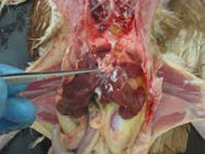
10. Retract the gastrointestinal tract to expose the reproductive tract
The spleen is present on the newly exposed, dorsal surface of the ventriculus. Sample the liver and spleen for fresh and fixed samples.Cut the colon distally. Remove the entire gastrointestinal tract and collect fixed samples of the duodenum and pancreas, jejunum, ileum, colon and caecum.
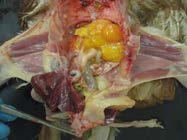
11. Anatomy after removal of the reproductive tract
If gut content is abnormal (very fluid, reddened etc), collect a fresh sample or swab.If toxicity is suspected, sample fresh proventricular and ventricular content.A fresh faecal or distal colon sample can be used to check for enteric parasites like coccidia.Sample the kidneys (fresh and fixed) and reproductive tract (fixed/fresh ovary and oviduct).
12. Gently peel the lungs from the rib cage
Grasp the caudolateral edge of the lung and apply traction medially. Incise or blunt dissect between the lungs and ribs to remove the lungs. Note the lungs are normally closely moulded to the ribs. Sample the lungs (fresh/fixed).
13. Open the crop and oesophagus
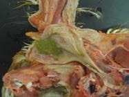
Check for exudates/ plaques that may indicate Trichomonas or Candida infection or hypovitaminosis A.Check the crop content as a clue to recent food intake.
14. Location of larynx
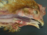
Examine the upper respiratory tract and alimentary tract.Examine the larynx and trachea for caseous exudate of infectious laryngeotracheitis (ILT). Sample both fresh and fixed trachea.Examine upper respiratory and alimentary tract for plaques (fowl pox).
15. Swab the sinuses for exudates
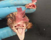
Remove the head with the secateurs. Cut across the upper beak/nares.Examine the sinuses for caseous exudate – swab for bacteria and/or Mycoplasma (chronic respiratory disease).
16. Brain exposed for sampling
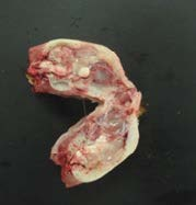
Skin the head and remove the lower beak. Split the skull to expose the brain by placing a heavy knife or cleaver on the midline.Swab or shell out half the brain for a fresh sample.Fix the skull with remaining brain for histopathology.
Remember: Sample lesions if not included in the samples mentioned above; fresh and fixed samples are ideal if lesions are large enough.
For more information, please contact the Animal Health Laboratories.