Calling a vet to investigate disease protects our marketsThe WA Livestock Disease Outlook provides information about recent livestock disease cases in Western Australia and diseases likely to occur in the next month. Calling a vet to investigate diseases when they occur provides surveillance evidence to our markets that we are free of reportable and trade-sensitive diseases. |
Recent livestock disease cases in WA
Abortions associated with bovine anaemia due to Theileria orientalis group (BATOG) in cows in the Great Southern
- In a mob of 12 Murray Grey cows between three and seven years old, five aborted full-term foetuses. No other clinical signs in the cows were reported.
- The local DPIRD field vet conducted an investigation and post-mortem on one aborted foetus. A full range of fresh and fixed tissues from the aborted foetus as well as blood samples and vaginal swabs from the affected cows were submitted for laboratory testing.
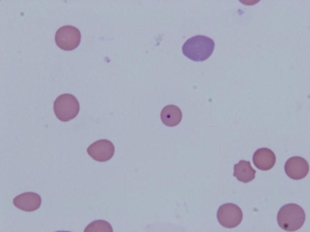
- Histopathology of the foetus was suggestive of foetal distress, with foetal atelectasis, and squames and meconium in the lung. The packed cell volume (PCV) was low in all of the cows tested, indicative of anaemia. Blood film examination revealed marked regenerative anaemia and organisms consistent with Theileria in all samples (see Image 1). Molecular testing was positive for T. orientalis Ikeda in all animals tested.
- Other infectious causes of abortion, including bovine pestivirus, vibriosis, leptospirosis and infectious bovine rhinotracheitis, as well as the reportable diseases bovine brucellosis, anaplasmosis, and babesiosis were ruled out in the investigation.
- Testing was subsidised as the results will contribute to demonstrating WA’s ongoing freedom from these trade-sensitive diseases.
- Read more about BATOG and the BATOG surveillance program in WA.

Image 1: Blood smear from one of the infected cows, showing sparse cellularity, erythrocytes containing piroplasms, poikilocytes and reticulocytes
Significant Disease Investigation program funds investigation into sudden death in Merino sheep in the Great Southern
- In a mob of 220 one-year-old, mixed-sex Merinos, 25 were found dead. Some sheep were down and convulsing before death. The sheep had been drenched one week prior.
- A private vet conducted an on-farm investigation and post-mortem, which was funded under the Significant Disease Investigation (SDI) program.
- Bloods, urine and a range of fresh and fixed tissues were submitted for laboratory testing, with provisional diagnoses of lead toxicity, listeriosis or a plant toxicity.
- Lead testing on the liver and kidney were below the level of detection, and bacterial culture, including specific Listeria cultures, was negative. A fluoroacetate assay on rumen contents was positive, supporting a diagnosis of fluoroacetate toxicosis.
- Plants in the Gastrolobium genus containing fluoroacetate (e.g. common box, heartleaf and narrow leaf poison bush) are likely to have highly toxic leaves at this time of year. Poisoning typically occurs when hungry stock gain access to bush or a new area containing the plants, and spring pasture dies and becomes less palatable.
- Subsidies under the SDI program are available for investigation of approved significant disease incidents in livestock and wildlife. See the SDI webpage for more information.
Lumpy skin disease ruled out in cattle in South-West
- Three 18-month-old Charolais cows from a mob of 300 had large wart-like skin lesions on the body (see Image 2). No other clinical signs were reported.
- Fresh and fixed tissue biopsies were submitted to DPIRD, with histopathology confirming fibropapillomas, and ruling out the reportable, lumpy skin disease.
- In cattle, fibropapillomas of the skin are due to infection with bovine papillomavirus 1, 2 or 5. In this case, the location of the lesion and morphology were consistent with BPV-2 infection, which is usually self-limiting and lesions regress within 12 months.
- Lumpy skin disease is a highly infectious disease of cattle caused by a poxvirus that is exotic to Australia. It is mostly transmitted by biting insects. Clinical signs include multiple, raised skin lesions, fever, salivation and nasal discharge.
- Lumpy skin disease is a priority disease due to its increasing international distribution and the major impact it would have on cattle productivity if it entered Australia.
