Recent news: exotic animal disease alerts
Lumpy skin disease
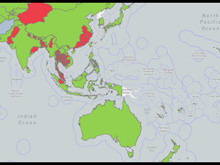
In March 2022, lumpy skin disease (LSD) was detected in Sumatra, Indonesia. The disease is continuing to expand in the East Asia and Pacific region, increasing the risk to Australia. See the exotic disease spotlight section below for more information on LSD, including clinical signs and transmission pathways.
Foot-and-mouth disease
In May 2022, foot-and-mouth disease (FMD) was detected in Java and Sumatra, Indonesia. Until this outbreak, Indonesia had been FMD free since 1986. FMD is a highly infectious viral disease that affects cloven-hoofed animals including sheep, cattle, pigs, goats, buffalo, camels, alpaca, llama and deer. Australia remains free of FMD, which increases our access to livestock and livestock product export markets.
Anyone keeping or working with cattle, sheep, goats or pigs should be aware of the signs of FMD: blisters on the mouth and drooling or limping animals. The most likely way foot-and-mouth disease could enter Australia is by feeding illegally imported meat and dairy products carrying the foot-and-mouth disease virus to pigs. For information on legal feed stuff for pigs, see the webpage: What you can and can’t feed pigs.
Anyone who sees livestock exhibiting any unusual signs is urged to call their local DPIRD veterinarian or Australia’s Emergency Animal Disease (EAD) Watch Hotline on 1800 675 888.
Japanese encephalitis virus update
Japanese encephalitis is an acute mosquito-borne viral disease that can cause reproductive losses and encephalitis in susceptible species. Pigs and horses are the most commonly affected animals, but the JE virus is zoonotic and can pose a serious risk to human health.
JE was detected in piggeries in Victoria, New South Wales, Queensland in February 2022 and South Australia in March 2022. There have been no detections of JE in Western Australia however vets are urged to be aware of the disease signs and to immediately report pigs or horses showing signs to your local DPIRD vet or the national Emergency Animal Disease Watch Hotline on 1800 675 888.
Signs in pigs include reproductive losses including abortions, mummified foetuses and stillborn or weak piglets. Tremors and convulsions are occasionally seen in pigs up to six months of age.
In horses, most clinical disease is mild and may be unrecognised, however fever, decreased or no appetite, lethargy, wobbliness and incoordination may occur. Some horses may die.
To enhance the detection of JE if it occurs in WA, vets can request access to subsidised investigations for clinically consistent horses and pigs.
DPIRD will provide $330 (GST inclusive) to private vets to collect an appropriate set of samples when JE is suspected. To qualify for the subsidy, veterinarians must have prior phone or email authorisation from their local DPIRD vet. DPIRD’s expectation is that you will subtract the $330 from the client’s invoice to encourage reporting.
More detail on JE in animals is available in the DPIRD JE webpage and the Outbreak website.
Recent livestock disease investigation
Foot-and-mouth disease ruled out in steers in the South-West
- Six out of 30 steers presented at a feedlot with salivation and mouth lesions. Another 20 steers arrived during the day from the same property of origin and ten of them were also salivating. On further inspection it was determined that half of the steers (25/50) had oral lesions.
- The steers had been purchased from a saleyard the day before, and there was no note of any unusual signs when the steers were loaded for transport.
- A private vet observed the clinical signs and called the EAD hotline to notify suspicion of the exotic animal disease FMD.
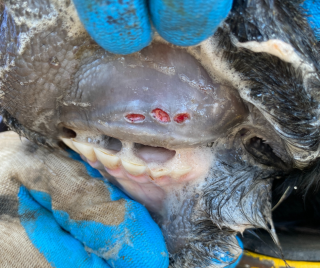
- A DPIRD vet attended the feedlot to examine and collect samples from the steers, noting rectal temperatures of 39°C–40°C and multiple ulcers on the buccal and the sublingual areas of affected cattle. No vesicles were detected, and no mucosal skin flaps were present. The steers were not lame, and no foot lesions were observed.
- Ensuring the use of correct personal protective equipment (PPE), the DPIRD vet collected complete blood samples, lesion swab samples in viral transport media and very small fresh gum biopsies from the edges of the lesions and submitted to the DPIRD laboratory for urgent testing.
- A second DPIRD vet attended the property of origin to examine cattle from the same cohort as the affected steers. Although there were no signs of salivation, on closer examination there were oral lesions on seven steers that were examined (Figure 1). Samples were submitted to the DPIRD laboratory for testing.
- There was no reported history that would identify a possible pathway for the introduction of any vesicular diseases.
- Samples were tested at DPIRD and were also sent to the Australian Centre for Disease Preparedness (ACDP, previously the Australian Animal Health Laboratory) for testing.
- Testing for the reportable diseases FMD, vesicular stomatitis, bovine viral diarrhoea Type II and malignany catarrhal fever returned negative results.
- Testing was positive for the parapox virus, bovine papular stomatitis virus, by electron microscopy and by PCR.
- Bovine papular stomatitis is a mild, self-limiting viral disease usually affecting cattle less than two years old. Infection with this virus usually shows no clinical signs in affected cattle, but when signs do occur they will typically manifest on the skin and mucous membranes, particularly around the mouth. People in close contact with infected animals may develop skin lesions and pustules on their hands, arms or legs.
- Clinical signs consistent with FMD should always be reported to a DPIRD vet or the Emergency Animal Disease hotline on 1800 675 888. Early detection supports faster and cheaper eradication.
Secondary photosensitisation in Merino ewes in the Midwest
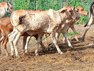
- In a flock of 550 mixed age Merino ewes, six had to be euthanised due to severe signs of jaundice, dehydration, facial swelling and scabbed oral lesions. More than 30 other ewes were mildly affected with swollen faces, jaundice and scabbing muzzle lesions (Figure 2).
- The producer contacted their local DPIRD vet to investigate the high morbidity and unusual signs of disease in their flock.
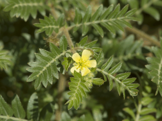
- The flock had moved onto a new pasture ten days earlier, which contained the weed caltrop (Figure 3). Caltrop is widespread throughout WA and can be identified by its star-shaped spiny burrs and small yellow flowers. Seeds remain dormant in the soil and germinate following summer rains.
- On post-mortem of one ewe, there was diffuse icterus of the carcase, bronchopneumonia and a swollen, yellow liver (Figure 4). Blood samples from three sheep, and faeces, fresh and fixed samples from one euthanised ewe were submitted with a provisional diagnosis of caltrop toxicosis and secondary photosensitisation.
- Histopathology revealed acute hepatocellular necrosis and acute renal tubular necrosis, both associated with formation of acicular clefts. This pattern is characteristic of steroidal saponin toxicosis, the hepatotoxic compound present in the weed caltrop.
- Testing of blood and liver samples excluded the exotic disease bluetongue virus. In the acute stage, sheep with bluetongue may present with pyrexia, salivation, nasal discharge causing crusting around the nostrils, swollen lips and tongue which may extend to include the ears, and petechial haemorrhages of the mucous membranes of mouth, nose and eyes.
- Although bluetongue virus is present in a monitored zone across northern Australia, bluetongue disease has never been reported in Australia. Suspicion of bluetongue virus disease must be reported immediately to your DPIRD vet or the EAD hotline on 1800 675 888.
- Other differential diagnoses in this case included the reportable diseases peste des petits ruminants, vesicular stomatitis and FMD. PCR and antibody testing was negative for all these diseases.
- The affected group was moved into a new paddock with no caltrop and plenty of shade. Livestock affected by photosensitisation should be protected from sunlight as it activates photosensitive substances in sparsely haired skin, which results in dermatitis and muzzle erosions. In the recovery period, access to high-protein feed should be limited.
- Results from this investigation help to provide evidence that WA is free from EADs and supports our access to markets.
In winter, watch out for these diseases
| Disease, typical history and signs | Key samples |
|---|---|
Arthritis in lambs
| Ante-mortem:
Post-mortem:
|
Copper deficiency in sheep and cattle
| Ante-mortem:
Post-mortem:
|
Bovine anaemia Theileria orientalis group (BATOG)
| Ante-mortem pre-treatment:
Post-mortem:
|
Note: Always include base samples and any clinical or gross lesions in submissions. For sample submission advice, contact your local DPIRD vet or the duty pathologist on +61 (0)8 9368 3351.
Register soon for your DPIRD regional private vet workshop
Don’t miss your chance to attend the regional private vet workshop in coming soon in August.
Topics covered will include:
- Updates on Indonesian situation with lumpy skin disease and Foot and Mouth disease
- Signs of LSD and FMD and what to do if you see these signs
- Japanese encephalitis in Australia
- Helping your clients manage the Johne’s disease risk
- Information on the emergency disease watch hotline
- Discussion of interesting cases by you, your local Field Vet and a DDLS Pathologist
- Case handling tips for suspect exotic animal diseases - precautions to avoid the possibility of spreading a disease
Participation is free to rural practitioners.
Contact your local DPIRD field vet to register your interest and to find out more details!
Exotic disease in the spotlight: lumpy skin disease
- Lumpy skin disease (LSD) virus is a Capripoxvirus that affects cattle and buffalo.
- LSD first occurred in Africa. From the late1980s it was detected in parts of the Middle East, from 2012 in Europe, and from 2019 in mainland South-East Asia, gradually moving east. In March 2022, it was detected in Singapore and Indonesia. LSD has never been recorded in Australia, but its presence in near neighbours has increased the likelihood of introduction.
- Bos taurus cattle are more susceptible to the disease than Bos indicus breeds, with some dairy breeds (Jersey, Guernsey, Friesian and Ayrshire) particularly susceptible. Asian water buffalo (Bubalis bubalis) can be infected, but clinical disease may be milder.
Clinical signs
- Typically presents as fever, multiple nodules on the skin and mucous membranes, which commonly become necrotic. Necrotic nodules may also occur within the alimentary system.
- Severity of clinical signs may vary from inapparent to severe, and disease signs in Bos indicus cattle may be difficult to see.
- Signs generally include a fever lasting 6-72 hrs, increased ocular, nasal and pharyngeal secretions, inappetence, milk drop, depression and reluctance to move. After 1–2 days nodules will erupt on the skin – these may cover the whole body or may be localised. The nodules will be raised, red and may be weeping. Regional lymph nodes will be enlarged, and regional dependent oedema may occur.
- If the respiratory tract is involved, respiratory distress may occur. Pneumonia is a common complication and may be fatal.
- The nodules may slough leaving full thickness lesions in the skin which commonly fill with purulent exudate. Scabs may also form on these nodules and may fall off, leaving large holes in the hide that can become infected.
- There is a rapid loss of condition, lesions may persist for 4-6 weeks and can take up to six months to fully resolve.
What to do if you see signs
Lumpy skin disease is a reportable disease in Australia. Eradication of LSD is difficult and early detection is essential for successful control and eradication. If you investigate a disease with these signs, contact your DPIRD vet or the emergency animal disease hotline on 1800 675 888.
More information on LSD can be found on our webpage.
Sampling guide for investigating skin lesions
DPIRD encourages and assists vets to investigate cases where livestock show signs similar to reportable diseases. Correct sampling increases the likelihood of a definitive diagnosis. Where possible, a full base sample set of blood (EDTA and plain tubes), fresh tissue and fixed tissues should be submitted. DPIRD’s livestock disease veterinary sampling guide provides guidance on general sample collection.
| Sample | Storage | Comments |
|---|---|---|
| Skin/mucous membrance (fresh and fixed) | Fixed can be held at room temperature, fresh samples should be chilled. | Fresh tissue biopsy preferred. Ensure samples are representative of lesions. For large lesions, sample interface with normal tissue and areas of different colour or consistency. See DPIRD’s histopathology sampling guide. |
| Vesicle fluid in viral transport media | Chilled | N/A |
| Swabs in transport media | Chilled | Submit swabs of mass and / or exudate. |
| Scabs or crusts | in plain container | N/A |
| Samples of relevant feed or plants (ergot etc.) | Chill fresh samples | If submitting hay samples, a minimum sample weight of 100 grams is required. Collect samples from up to 10 bales, up to 1kg total weight. |
| Maggots | Place maggots in boiled water for 5-10 seconds until they appear white and float, then place into alcohol (70% or higher). See the Northern Australian Biosecurity Surveillance network guide for collecting maggots for more information. | Ensure clean sampling equipment is used and individual specimens stored separately to prevent DNA cross contamination. If possible, provide a photo in situ. |
| Photos of skin lesions | N/A | N/A |
| Blood | Plain and EDTA blood tubes | For PCR or ELISA if required. |
| Additional base sample set | Variable | See DPIRD’s livestock disease veterinary sampling guide for full list. |
WA Livestock Disease Outlook highlights the benefits of surveillance
Australia’s ability to sell livestock and livestock products depends on evidence from our surveillance systems that we are free of particular livestock diseases. The WA livestock disease outlook – for vets summarises recent significant disease investigations by DPIRD vets and private vets that contribute to that surveillance evidence.
Feedback and subscriptions
We welcome feedback. To provide comments or to subscribe to the monthly email newsletter, WA livestock disease outlook, email waldo@dpird.wa.gov.au


