Vet workshop supports WA's animal health status
34 rural and peri-urban veterinarians participated in a workshop coordinated by DPIRD last month.
The Livestock Disease Investigation Workshop provided updated information on WA’s surveillance systems, reinforced how the daily work of vets contributes to protecting WA’s livestock markets, and aimed to improve vets’ ability to detect emergency animal diseases quickly.
Topics for discussion this year included the Wooroloo bush fire response, practical tips for getting vets on farm and the African swine fever disease investigation in Laos.
An interactive epidemiology session was a popular activity on the day, with vets breaking into groups to conduct a mock outbreak investigation. Attendees particularly enjoyed the wide range of topics covered during the workshop.
A practical post mortem session was also held for 17 mixed practice vets. The post mortem session was led by a DPIRD senior pathologist, and participants had the opportunity to practice post mortem sampling and brain removal technique with assistance.
Recent livestock disease investigations
Listeriosis diagnosed in ewe
- A single ewe was noticed circling and with a muted response to stimuli.
- A producer contacted a DPIRD field veterinary officer who visited the property to conduct a disease investigation.
- The vet examined the affected ewe, which displayed a severe bilateral horizontal nystagmus.
- On post-mortem there was a severe diffuse reddening of the lungs. No gross abnormalities of the brain were recorded.
- The vet submitted a full set of fixed and fresh samples, including brain, to DPIRD Diagnostics and Laboratory Services (DDLS) for analysis.
- Investigations of neurological disease in sheep and cattle may be eligible for subsidies under the transmissible spongiform encephalopathy (TSE) program. In this instance the ewe was under 18 months so did not meet the criteria for the TSE program. However, the cost of the investigation and the laboratory testing was covered by DPIRD as part of encouraging reporting and investigation of unusual disease signs in WA livestock.
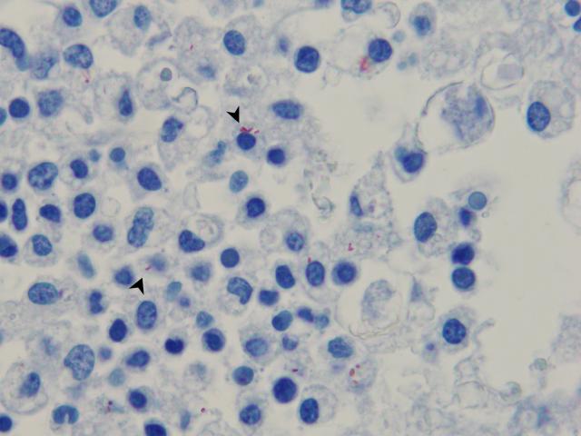
- Histopathological examination of the brain tissue led to a diagnosis of suppurative encephalitis and malacia affecting the brainstem. Listeria immunohistochemistry stained plump rods within the lesions. An example of listeria bacteria is shown in Figure 1, from a separate case of listeriosis in a cow.
- Bronchopneumonia was also diagnosed on histopathological examination of the lungs.
- The lung tissue was cultured and there was a moderate growth of the bacteria Mannheimia haemolytica.

Sudden death and neurological signs in feedlot cattle
- 6 out of 120 weaner cattle died suddenly in a feedlot, with a further 5 affected with neurological signs.
- A private vet attended the property and accessed a significant disease investigation subsidy by phoning his local DPIRD field veterinary officer for approval.
- Two clinical animals were examined by the visiting vet, both in lateral recumbency and unable to rise. The vet collected blood from both of these animals prior to euthanasia and then conducted a post-mortem examination. A full range of fixed and fresh samples was submitted to DPIRD laboratory.
- On post-mortem, one of the weaners had no obvious gross lesions, while the other had patchy discolouration of the lungs and fibrinous adhesions between the lungs and the thoracic wall (pleurisy).
- Histopathological analysis of the brain tissue at the laboratory led to a diagnosis of thromboembolic meningoencephalitis caused by Histophilus somni infection. Thromboembolic lesions were also seen in some heart, lung and muscle tissues on histopathology.
- Histophilus somni was cultured from the brain tissue of both animals and from the lung in one animal.
- Histophilosis is frequently found in feedlot situations when environmental stressors and pathogenic strains of the bacterium combine.
- Lead toxicity was also excluded in this case. Lead ingestion by food-producing animals presents a risk to human food safety and access to export markets.
- It is important to investigate neurological disease or sudden deaths in livestock, to ensure they are not due to an exotic disease with similar signs. Investigations may be eligible for subsidy under a number of surveillance incentives.
- For more information or advice on conducting livestock post-mortems or sampling required for disease exclusions, contact your local DPIRD field veterinary officer or visit the livestock disease veterinary sampling guide.
In early winter, watch for these livestock diseases
| Disease, typical history and signs | Key samples |
| Pregnancy toxaemia in ewes
| Ante-mortem
Post-mortem
|
| Calf scours
| Ante-mortem
Post-mortem
|
Livestock disease investigation kits for private vets
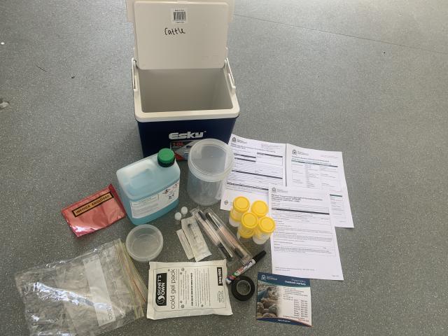
DPIRD field vets are distributing livestock disease investigation kits. The kits contain sampling equipment to aid in post-mortem sample collection. Kit contents include: hard esky for sending samples to DPIRD Diagnostics and Laboratory Services (DDLS), various pots for fixed and fresh tissues, formalin, laboratory forms, ice brick and swabs. Empty eskies will be collected from DDLS and restocked by your local field vet.
Contact your local field vet if you would like to receive kits for your clinic. There is no cost involved.

