Livestock disease investigations protect our marketsAustralia’s ability to sell livestock and livestock products depends on evidence from our surveillance systems that we are free of particular livestock diseases. The WA livestock disease outlook – for vets summarises recent significant disease investigations by Department of Primary Industries and Regional Development (DPIRD) vets and private vets that contribute to that surveillance evidence. |
Recent livestock disease cases in WA
Several cases of annual ryegrass toxicity (ARGT) cause livestock deaths
- Since 1 March 2018, DPIRD has investigated 16 cases of ARGT in livestock in Western Australia, including seven cases in cattle, six in sheep, two in horses and one goat.
- In one case, a herd of 50 two-year-old Jersey cows was affected, with one death and one cow affected by sudden onset of ataxia, inappetence, muscle fasciculations and recumbency. The herd was fed silage and grazing a mixed pasture.
- Given that the clinical signs and deaths were unusual, DPIRD granted a private vet a subsidy of $330 under the Significant Disease Investigation program.
- Samples including faeces, blood, pasture, silage, and a range of fresh and fixed tissues were submitted to the DPIRD laboratory with provisional diagnoses including ARGT, listeriosis and thrombotic meningoencephalitis.
- Both the pasture and faecal samples tested positive for ARGT. Clinical biochemistry indicated a mild increase in hepatobiliary parameters (GGT, GLDH), which is often noted in ARGT. Marked increases in CK and ALT were also present, indicating muscle injury likely due to recumbency and/or misadventure while ataxic. Histopathology did not detect significant lesions.
- ARGT at this time of year is more likely to be from affected supplementary feed such as hay or silage. ARGT on pasture is most often seen in spring when livestock graze pastures containing infected ryegrass seed heads. Note that drying the grasses for hay does not eliminate the toxin.
- See DPIRD’s recent media release on ARGT, as well as the DPIRD ARGT webpage for more information.
Bluetongue and screw-worm fly excluded in sheep at abattoir
- In the past month, DPIRD has received two submissions to exclude reportable diseases from sheep at abattoir.
- In the first case, five Merino ewes from a line of 1044 had a nasal discharge, conjunctivitis and corneal opacity. Histopathology and biochemistry at DPIRD showed chronic keratitis and blepharitis, leading to a diagnosis of infectious keratoconjunctivitis. Given that bluetongue virus also causes nasal discharge in sheep, a bluetongue PCR and ELISA were also run at DPIRD, both of which were negative.
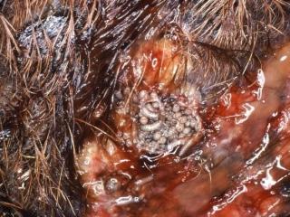
- In the second case, a Merino ewe with a maggot-infested wound was identified at ante-mortem inspection. The ewe was removed from the slaughter line, euthanased and maggots were collected into 70% ethanol and submitted to DPIRD to exclude the exotic screw-worm fly (see Figure 2). The larvae were identified as sheep blowfly, Lucilia cuprina. See the DPIRD flystrike tools webpage.
- The results of these cases contribute to demonstrating WA’s and Australia’s freedom from these diseases, which gives us a market advantage and allows us to export livestock products widely.
- Always report any suspicion of a reportable disease immediately to a DPIRD field vet or the Emergency Animal Disease hotline on 1800 675 888. Early detection = faster eradication.
Infectious laryngotracheitis and chronic respiratory disease in mixed age hens
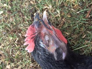
- In a flock of 100 mixed-age birds, including turkeys, pheasants and chickens, eight chickens died suddenly over a two-week period. Several birds were affected by ocular swelling and discharge (see Figure 2).
- Two birds were submitted to the DPIRD laboratory for investigation. On post-mortem examination and histopathology, they had a severe tracheitis with intranuclear inclusions, moderate to severe bronchopneumonia and chronic sinusitis. Virology of tracheal swabs revealed an infection with infectious laryngotracheitis (Gallid herpesvirus 1), and culture for Mycoplasma species (both M. gallisepticum and M. synoviae) was positive for both birds indicating the flock was concurrently affected by chronic respiratory disease (CRD).
- Reportable diseases including avian influenza and Newcastle disease were ruled out as the cause of the disease.
- DPIRD has a guide to assist vets in investigating poultry disease, including how to conduct a post-mortem and samples to collect to maximise the likelihood of reaching a diagnosis.
In early autumn, watch for these livestock diseases
| Disease, typical history and signs | Key samples |
|---|---|
Grain overload in stock
| Post-mortem:
|
Pink eye in cattle and sheep
| Ante-mortem:
|


